


How Intraoral Scanners Work

Principle / How Intraoral Scanners Work
Overview of the basic scanning process used by intraoral scanners:
The basic scanning process used by intraoral scanners
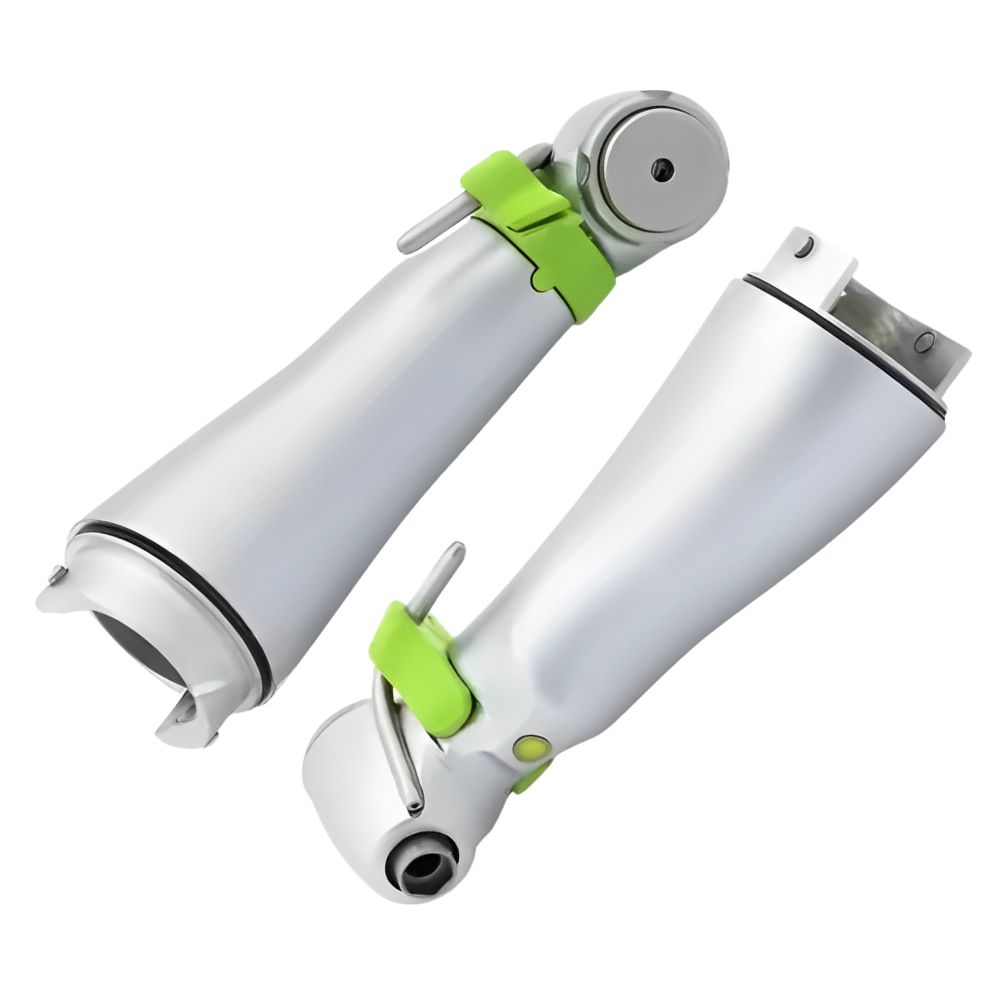
Intraoral scanners use a handheld wand that is moved around the mouth to capture images of the teeth and surrounding tissues. The tip of the scanner wand contains optic components including:
what is intraoral scanner wikipedia
- One or more cameras to capture images
- Laser, structured light, or other illumination source
- Lenses, mirrors and sensors to measure distortions
As the scanner tip is slowly dragged along the teeth, it projects a laser or structured light pattern onto the surfaces while rapidly capturing images. The scanning software analyzes the pattern distortions picked up by the sensors to map out the contours and textures of the teeth in 3D.
Hundreds or thousands of images are taken from slightly different angles as the scanner moves around the mouth. Advanced processing algorithms analyze this image stream data to stitch and blend the images together into a seamless 3D model.
The scanner software accounts for any irregularities in movement or positioning, using accelerometer and gyroscope data to align the images properly. This allows even novice users to get accurate scans without needing perfectly steady hand motions.
Once fully processed, the scan data file can be exported as an open STL file or proprietary file format. CAD software then allows the 3D model to be used for various applications like creating surgical guides, crowns, aligners and more.
So in summary, intraoral scanners use a wand to capture a video-like stream of images that are automatically converted by sophisticated software into a detailed 3D model of the oral anatomy. This digital impression can then be utilized for a variety of dental treatments and appliances.
Basic scanning patterns
Basic scanning patterns used with intraoral scanners:
To fully capture the teeth and oral anatomy, the scanner wand must be moved around the mouth in a careful, methodical pattern. Proper wand motion is important to effectively stitch together the many images into an accurate model.
For upper teeth, it’s recommended to start scanning from the posterior teeth and slowly progress forward. The wand tip should follow the curve of the arch, staying in close contact with the teeth and just slightly angled toward the occlusal plane.
For lower arches, the same posterior-to-anterior pattern is used, scanning the lingual side of the teeth. The wand is inverted but still held at a slight angle toward the occlusal.
The motion should be slow, smooth, and steady as the scanner acquires a constant stream of images. Abrupt movements or lifting the scanner off the teeth can disrupt the scanning process.
The wand is like a video camera, capturing frames constantly from every vantage point. So overlapping scanning from multiple angles helps improve detail and accuracy. Difficult-to-reach areas may require special positioning.
Bite registration requires holding the wand still while the patient closes into occlusion, to link the upper and lower dental arches together. Motionless scanning may also be used for small isolated areas.
With practice, the scanning patterns become second nature. While software can compensate for imperfections, proper technique is key for the most accurate digital impressions.
How powders and opacifying agents are used with intraoral scanners:
-
Reducing Reflectivity
- Enhancing Surface Detail
- Improving Scanning Efficiency
- Patient Comfort
- 5. Preventing Fogging
Projecting light/lasers and capturing the distortion with sensors
- Light Projection
Intraoral scanners often use structured light or laser projection systems to illuminate the surfaces being scanned. Structured light involves projecting a known pattern of light onto the dental structures. Lasers, which emit coherent and focused beams of light, are also commonly employed.
- Pattern Deformation
- Capture by Sensors
- Triangulation and Depth Calculation
- Real-time Processing
- Creation of Digital Models
Advantages of using light or lasers in intraoral scanners include:
-
Accuracy
-
Speed
-
Non-invasiveness
- Real-time Feedback
Generating multiple images from different angles
- Multiple Cameras or Light Sources
- Structured Light or Laser Projection
- Simultaneous Image Capture
- Coordinate Alignment
- Real-time Processing
- Continuous Scanning
- Feedback and Visualization
- Comprehensive Coverage
- Enhanced Accuracy
- Efficiency
- Better Visualization
Conversion of images into 3D rendering by software
- Image Acquisition
- Feature Extraction
- Correspondence Matching
- Triangulation
- Surface Reconstruction
- Texture Mapping (Optional)
- Post-Processing and Refinement
- Visualization
This process is widely used in various applications, from reconstructing 3D models of objects for virtual reality to generating anatomical models from medical imaging data. The specific algorithms and techniques employed can vary based on the application and the characteristics of the input data.
Stitching images together into a complete model.
- Image Alignment
- Feature Matching
- Homography Estimation
- Image Warping
- Blending
- Global Optimization (Optional)
- Post-Processing
- Output
Why do scanners often use white or blue light for accuracy?
- Optimal Scattering Properties
- Improved Depth Perception
- Reduced Reflection and Glare
- Color Differentiation
- Less Heat Generation
- Compatibility with Optical Sensors
- Clinical Considerations
IR cameras and gyroscope/accelerometer data were utilized.
- IR Cameras
- Gyroscope and Accelerometer Data
- Dynamic Image Registration
- Reduction of Artifacts
- Improved User Experience
Stereophotogrammetry triangulation process
- Image Acquisition
- Calibration
- Feature Matching
- Epipolar Geometry
- Triangulation
- Bundle Adjustment (Optional)
- Generation of 3D Model
- Texture Mapping (Optional)
Steps for calibrating the scanning wand before use.
- Power On the Scanner
- Prepare Calibration Tool or Target
- Place the Calibration Tool in the Field of View
- Initiate Calibration Mode
- Follow on-screen Instructions
- Capture Calibration Images
- Analysis and Adjustment
- Verify Calibration Accuracy
- Document Calibration Results
- Regular Calibration Checks
- Calibration Quality Assurance
Frequently Asked Questions (FAQs) about How Intraoral Scanners Work:
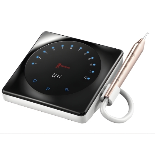
An intraoral scanner is a device used by dentists to capture detailed 3D images of a patient’s teeth and oral structures.
Intraoral scanners use advanced optical technology to capture thousands of images per second while scanning the inside of a patient’s mouth. These images are then stitched together to create a precise 3D model.
- Typical components include a handheld wand with a camera, a light source, and software to process the captured images.
Intraoral scanners eliminate the need for traditional dental impressions, which can be uncomfortable for patients. They also provide highly accurate digital models for various dental procedures, such as crowns, bridges, and aligners.
Yes, intraoral scanners are safe to use. They are designed to be non-invasive and do not emit any harmful radiation.
The duration of a scan can vary depending on the complexity of the case, but a typical scan usually takes a few minutes to complete.
While intraoral scanners are incredibly versatile, they may not be suitable for every dental procedure. Your dentist will determine the best approach based on your specific needs.
Coverage for intraoral scanners may vary depending on your dental insurance plan. It’s best to check with your provider to determine coverage.
Intraoral scanners should be properly cleaned and maintained according to the manufacturer’s instructions to ensure optimal performance and longevity.
Intraoral scanners are known for their high level of accuracy, often providing measurements within microns of precision.