


Intraoral scanners – PRESENT and future
A Broad Overview of Intraoral Scanning
The Evolution of Intraoral Scanning Technology
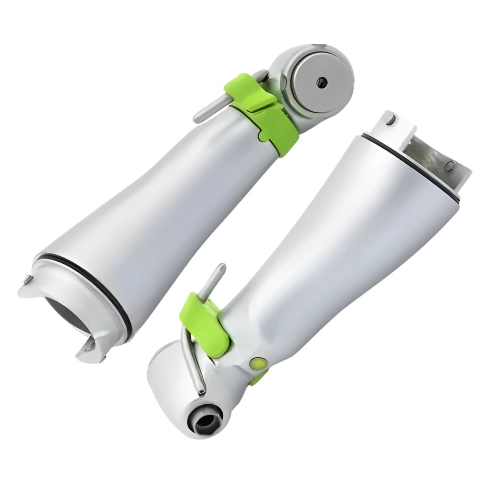
Intraoral Scanner Basics and Methodology
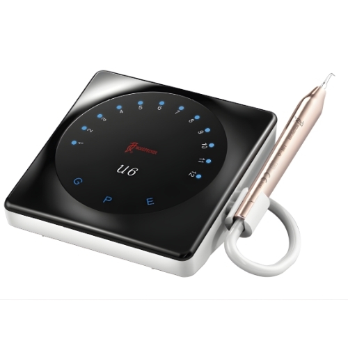
Intraoral Scanner Options and Differences
Clinical Advantages of Intraoral Scanners
Limitations and Considerations of Intraoral Scanners
Patient Perspectives on Intraoral Scanning
Effect of Intraoral Scanners on Dental Laboratories
Implementation Costs and Economics
The Future of Intraoral Scanners
Conclusion
Frequently Aske Questions
. What is an intraoral scanner and how does it work?
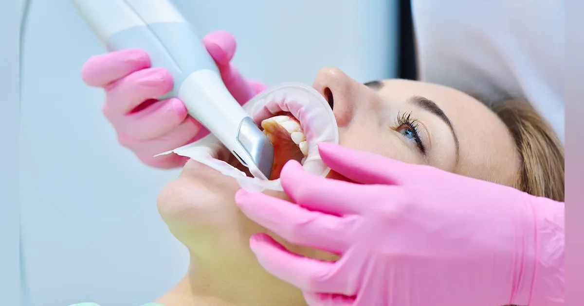
An intraoral scanner is a handheld, wand-like digital device that acts like a 3D camera for the mouth. It uses safe structured light or lasers to capture precise images of your teeth and gums. As the dentist moves it around your mouth, sophisticated software stitches these images together into a complete, accurate 3D digital model, eliminating the need for traditional, messy impression putty and trays.
2. Is an intraoral scan more comfortable than a traditional impression?
Absolutely. For most patients, especially those with a strong gag reflex or dental anxiety, it’s a vastly more comfortable experience. The process is quick, there’s no unpleasant taste or feeling of being choked by a tray full of putty, and you can breathe and swallow normally throughout the scan.
3. What are the main benefits for my dental treatment?
The benefits are significant. Digital scans are extremely accurate, leading to better-fitting crowns, veneers, and aligners with fewer adjustments. The process is faster, often shortening your chair time. It also enables advanced treatment planning, allowing your dentist to show you a preview of potential results and plan procedures like implant placement with great precision.
4. Are there any downsides or limitations to intraoral scanning?
The technology is excellent but has some considerations. It requires a dry field, so controlling saliva is important. Scanning shiny surfaces like metal crowns or implant parts can sometimes be challenging. There’s also a significant upfront cost for the dental practice, and the dentist and staff need training to use the system proficiently.
5. How does intraoral scanning affect the dental lab and my restoration?
It creates a more efficient and accurate digital workflow. Your scan is sent electronically to the lab in seconds, eliminating shipping delays and potential damage to physical models. Labs use this digital file to design and often mill or 3D-print your restoration with high precision, facilitating better communication and collaboration with your dentist.
6. Will intraoral scanners make physical impressions completely obsolete?
While traditional impressions are becoming less common, they aren’t entirely obsolete yet. Very complex cases or specific situations (like severe moisture control issues) might still use them. However, intraoral scanning is undoubtedly becoming the new standard of care due to its advantages, and its use will continue to grow as the technology becomes more accessible and capable.