


Major Types of Scanning Technologies
Overview of some of the major types of scanning technologies used in intraoral dental scanners:
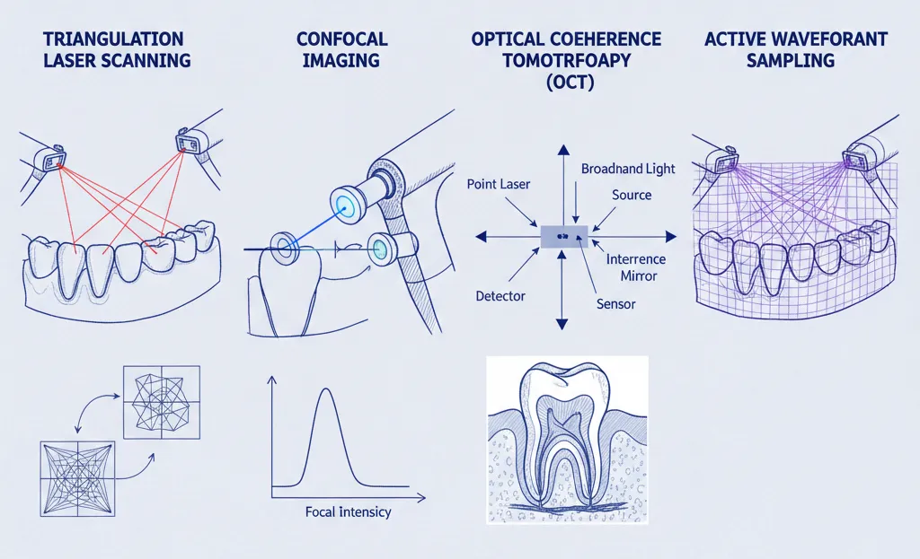
Triangulation Laser Scanning
- Uses a laser light source and 1 or 2 cameras at known angles to scan the teeth
- A laser stripe is projected on the surface, and distortions are analyzed
- Allows precise, fast scanning with good depth information
- Limited by line-of-sight views and struggles with dark surfaces
Triangulation Laser Scanning in Intraoral Scanners: A Technological Marvel in Dentistry
Triangulation laser scanning is a pivotal technology in the realm of intraoral scanners, revolutionizing the way dental impressions are taken. This essay explores the intricacies of triangulation laser scanning, its historical development, components, advantages, applications in dentistry, challenges, and future prospects.
Basics of Triangulation Laser Scanning:
At its core, triangulation laser scanning employs fundamental principles of triangulation to capture three-dimensional data. In the context of intraoral scanners, this involves the utilization of laser beams, optics, and detectors to create highly accurate digital impressions of the oral cavity.
Historical Context:
Early attempts at 3D scanning in dentistry paved the way for the evolution of triangulation laser scanning technology. Pioneering devices and innovations marked significant milestones in the development of this technology.
Key Components:
Triangulation laser scanning in intraoral scanners involves crucial components such as the laser source, optics and lenses, and detector systems. The type of lasers used, their wavelengths, and the role of optics and detectors play a crucial role in achieving precision and accuracy.
Advantages:
Triangulation laser scanning boasts several advantages, including high precision and accuracy, rapid scanning speed, enhanced detail capture, and reduced sensitivity to environmental factors. These qualities make it a preferred choice in various dental applications.
Applications in Dentistry:
The versatility of this laser scanning is evident in its applications across different dental fields. From restorative dentistry for crown and bridge design to orthodontics for aligner fabrication and prosthodontics for denture design, this technology has wide-ranging implications.

Challenges and Limitations:
Despite its merits, triangulation laser scanning faces challenges such as cost and accessibility, a learning curve for dental professionals, and limitations in the field of view and scanning depth.
Integration with Other Technologies:
Triangulation laser scanning seamlessly integrates with other technologies like CAD/CAM systems, 3D printing, and augmented reality, enhancing its utility in comprehensive treatment planning and execution.
Case Studies and Success Stories:
Examining notable examples of triangulation laser scanning implementation reveals improved clinical outcomes, heightened patient satisfaction, and widespread acceptance within the dental community.
Future Developments and Innovations:
The future of triangulation laser scanning in intraoral scanners holds promise, with ongoing efforts focused on miniaturization, integration with artificial intelligence, and potential applications in emerging fields like tele-dentistry.
Conclusion:
Triangulation laser scanning stands as a cornerstone in the evolution of intraoral scanners, offering unparalleled precision and versatility in dental applications. As technology continues to advance, the transformative impact of this technology on dentistry is poised to grow, ushering in a new era of digital oral healthcare.
Confocal Imaging
- Employs a point laser source and objective lens with a pinhole filter
- Measures intensity of reflected light at different focal depths
- Excellent at capturing details on uneven surfaces
- Limitations include slow scan speed and shallow depth of field
Confocal Imaging Technology in Intraoral Scanners: Shaping Precision Dentistry
Introduction: Confocal imaging technology has emerged as a groundbreaking method in intraoral scanners, providing dental professionals with a powerful tool for capturing high-resolution three-dimensional images of the oral cavity. This essay delves into the principles, components, advantages, applications, and potential future developments of confocal imaging technology in the field of dentistry.
Basics of Confocal Imaging: Confocal imaging involves the use of a focused beam of light to capture images with enhanced clarity and depth. In intraoral scanners, this technology employs a confocal laser scanning system to precisely measure the reflected light, allowing for the creation of detailed digital impressions.
Historical Context: The incorporation of confocal imaging technology in dentistry represents a significant advancement in the quest for accurate and efficient oral imaging. This section explores the historical development and key milestones leading to the integration of confocal imaging into intraoral scanners.
Key Components: Confocal imaging technology in intraoral scanners comprises essential components such as a laser light source, a pinhole aperture, and a detector. The focused laser beam interacts with dental surfaces, and the pinhole aperture selectively filters the reflected light, resulting in improved resolution and contrast in the captured images.
Advantages of Confocal Imaging: Confocal imaging technology offers several advantages in intraoral scanning, including exceptional depth resolution, reduced image distortion, and the ability to capture fine details. These advantages contribute to enhanced accuracy and efficiency in various dental applications.
Applications in Dentistry: The versatility of confocal imaging extends across multiple dental disciplines. In restorative dentistry, it facilitates precise impressions for crowns and bridges. In orthodontics, it aids in digital models for treatment planning, and in prosthodontics, it plays a crucial role in the design and fabrication of prosthetic restorations.
Challenges and Limitations: While confocal imaging technology has transformative potential, it faces challenges such as cost, complexity, and a learning curve for dental professionals. Understanding and addressing these challenges is crucial for widespread adoption in dental practices.
Integration with Other Technologies: Confocal imaging seamlessly integrates with other digital technologies like CAD/CAM systems and 3D printing, offering a comprehensive solution for digital dentistry. This section explores the synergies between confocal imaging and other cutting-edge dental technologies.
Case Studies and Success Stories: Examining real-world applications of confocal imaging in intraoral scanners reveals success stories, demonstrating improved clinical outcomes, enhanced patient experiences, and streamlined workflows in dental practices.
Future Developments and Innovations: The future of confocal imaging in intraoral scanners holds exciting possibilities. Ongoing research focuses on miniaturization, increased automation, and integration with artificial intelligence, paving the way for more accessible and advanced dental imaging solutions.
Conclusion: Confocal imaging technology stands at the forefront of precision dentistry, offering a paradigm shift in how intraoral scanners capture and reproduce detailed images of the oral environment. As this technology continues to evolve, its impact on dental diagnostics and treatment planning is set to grow, ushering in a new era of digital excellence in oral healthcare.
Optical Coherence Tomography (OCT)
- Uses a broadband light source and interferometry to gather scans
- Light is split into reference and sample beams to measure echoes
- Provides very detailed, high-resolution subsurface images
- Slow capture rate makes it difficult to scan full arches quickly
Optical Coherence Tomography (OCT) in Intraoral Scanners: Advancing Precision Imaging in Dentistry
Introduction: Optical Coherence Tomography (OCT) has emerged as a cutting-edge technology in intraoral scanners, offering dental professionals a non-invasive and high-resolution imaging solution for comprehensive diagnostics. This essay explores the fundamental principles, components, advantages, applications, challenges, and future prospects of OCT technology in the context of intraoral scanners.
Basics of Optical Coherence Tomography: OCT utilizes low-coherence interferometry to capture detailed cross-sectional images of biological tissues with micron-level resolution. In intraoral scanners, this technology enables the visualization of internal structures of teeth and surrounding tissues, providing valuable diagnostic information.
Historical Context: The integration of OCT into intraoral scanners represents a significant milestone in the evolution of dental imaging. This section explores the historical development of OCT technology and its gradual integration into the field of dentistry.
Key Components: OCT technology in intraoral scanners comprises key components, including a low-coherence light source, a beam splitter, a reference mirror, and a detector. The interference pattern generated by the interaction of light from the sample and reference arms is analyzed to produce detailed three-dimensional images.
Advantages of OCT Imaging: OCT brings several advantages to intraoral scanning, including high resolution, non-invasiveness, real-time imaging capabilities, and the ability to visualize subsurface structures. These advantages contribute to improved diagnostic accuracy and treatment planning.
Applications in Dentistry: The applications of OCT in dentistry are diverse. In restorative dentistry, it aids in detecting early carious lesions and assessing restoration integrity. In periodontics, OCT provides insights into periodontal and gingival tissues, and in endodontics, it assists in visualizing root canal anatomy.
Challenges and Limitations: Despite its promising capabilities, OCT technology in intraoral scanners faces challenges such as cost, limited penetration depth, and the need for specialized training. Addressing these challenges is crucial for wider acceptance and integration into routine dental practice.
Integration with Other Technologies: OCT seamlessly integrates with other digital technologies, such as CAD/CAM systems, enabling a more comprehensive approach to digital dentistry. The synergies between OCT and other technologies enhance the overall diagnostic and treatment capabilities in dentistry.
Case Studies and Success Stories: Examining case studies and success stories highlights the practical applications and positive outcomes of OCT in intraoral scanners. These examples showcase the technology’s effectiveness in improving diagnostic accuracy and patient outcomes.
Future Developments and Innovations: The future of OCT in intraoral scanners holds exciting possibilities. Ongoing research focuses on enhancing device portability, increasing scanning speed, and integrating artificial intelligence for automated analysis, paving the way for more efficient and accessible dental imaging solutions.
Conclusion: Optical Coherence Tomography (OCT) has become a cornerstone in the realm of intraoral scanners, offering unparalleled insights into the internal structures of the oral cavity. As this technology continues to advance, its integration into routine dental practice is poised to bring transformative changes, ensuring precision and excellence in oral healthcare diagnostics and treatment planning.
Active Wavefront Sampling
- Projects a changing pattern of multiple rays onto the teeth
- Analyzes the deformation of ray pattern detected by sensors
- Generates accurate 3D models with good contrast and speed
- More technique-sensitive than passive scanning methods
Active Wavefront Sampling Technology in Intraoral Scanners: Elevating Precision and Speed in Dental Imaging
Introduction: Active Wavefront Sampling (AWS) technology stands as a pioneering force in intraoral scanners, providing a dynamic and rapid approach to digital impression capture in dentistry. This essay explores the principles, components, advantages, applications, challenges, and future potential of AWS technology in the context of intraoral scanners.
Basics of Active Wavefront Sampling: AWS technology involves the active projection of structured light patterns onto the dental surface. By analyzing the deformation of these patterns, the intraoral scanner rapidly generates a high-resolution three-dimensional representation of the oral structures. This method significantly improves the speed and precision of digital impressions.
Historical Context: The integration of AWS technology into intraoral scanners represents a significant leap forward in the evolution of digital dental imaging. This section explores the historical development of AWS and its gradual adoption in the dental field.
Key Components: AWS technology in intraoral scanners consists of essential components, including a light projector, a camera system, and sophisticated algorithms for real-time data processing. The synergy of these components allows for the accurate reconstruction of the dental anatomy.
Advantages of Active Wavefront Sampling: AWS brings forth several advantages, such as rapid image acquisition, enhanced accuracy, and the ability to capture intricate details in real time. The dynamic nature of AWS makes it particularly valuable in time-sensitive dental procedures.
Applications in Dentistry: The applications of AWS in dentistry are diverse. In restorative dentistry, it aids in the precise capture of tooth preparations for crowns and bridges. In orthodontics, AWS facilitates the swift generation of digital models for treatment planning, enhancing overall efficiency in clinical workflows.
Challenges and Limitations: While AWS technology offers remarkable advantages, challenges include the potential sensitivity to ambient lighting conditions and the need for continuous calibration. Addressing these challenges is essential for optimizing the reliability of AWS in various clinical settings.
Integration with Other Technologies: AWS seamlessly integrates with other digital technologies, including CAD/CAM systems, enabling a streamlined approach to comprehensive digital dentistry. The compatibility of AWS with various digital platforms enhances its versatility in treatment planning and execution.
Case Studies and Success Stories: Reviewing case studies and success stories provides insights into the practical applications and positive outcomes of AWS in intraoral scanners. These examples demonstrate how AWS contributes to improved clinical precision and patient satisfaction.
Future Developments and Innovations: The future of AWS in intraoral scanners holds exciting prospects. Ongoing research focuses on refining algorithms, improving portability, and exploring the potential integration with artificial intelligence, paving the way for more advanced and user-friendly dental imaging solutions.
Conclusion: Active Wavefront Sampling technology has emerged as a driving force in the evolution of intraoral scanners, redefining the landscape of digital impressions in dentistry. As AWS continues to evolve, its integration into routine dental practice is poised to bring about transformative changes, ensuring unparalleled precision and efficiency in oral healthcare diagnostics and treatment planning.
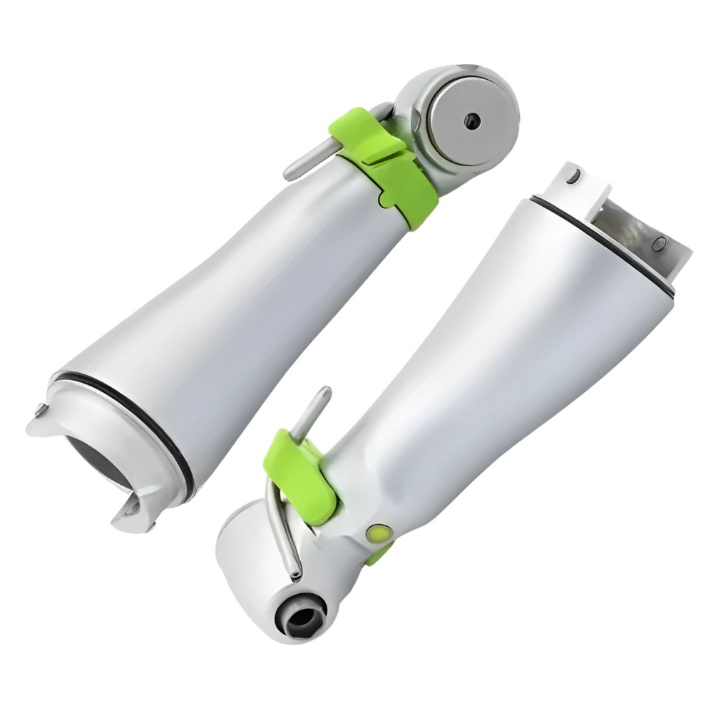
If you are looking for quality dental repair parts, read this article
RESUME:
While each technology has pros and cons, the optimal approach depends on the desired scanning performance and application. Continued development aims to combine strengths and minimize limitations.
Triangulation Laser Scanning is prized for its high precision and rapid scanning speed. It uses a laser stripe and angled cameras to create accurate 3D models with good depth information, making it excellent for fast, detailed captures of tooth preparations for crowns, bridges, and digital models.
Confocal Imaging excels at capturing exceptional detail on uneven surfaces. It uses a point laser and a pinhole filter to measure reflected light at different focal depths, providing high-resolution images with reduced distortion. This makes it ideal for capturing fine marginal details crucial for restorative and prosthetic work.
Unlike surface-scanning technologies, OCT provides detailed, high-resolution subsurface images. It uses interferometry to visualize internal tooth structures and surrounding tissues at a micron level. This non-invasive capability is revolutionary for early caries detection, assessing restoration integrity, and evaluating periodontal and gingival health.
Active Wavefront Sampling (AWS) achieves rapid image acquisition by projecting and analyzing a changing pattern of multiple light rays onto the teeth. By dynamically processing the deformation of this pattern in real-time, it generates accurate 3D models quickly, which is highly beneficial for efficient, time-sensitive clinical workflows.
A frequent trade-off is between scanning speed, depth of field, and technique sensitivity. For instance, Confocal Imaging can be slower with a shallow depth of field, while OCT has a slow capture rate for full arches. Triangulation Laser and AWS can be limited by line-of-sight or ambient lighting. The “optimal” technology often depends on the specific clinical application and desired